
Software Vendors & OEMs
Ready to integrate web DICOM PACS Viewer and connectivity solutions

CARDIOLOGY & ULTRASOUND
DICOM VIEWER
MedDream DICOM ECG & US Viewer provides standard Cardiology image manipulation tools, but also a way to read, manipulate and interpret electrocardiography data. Cardiology & Ultrasound Viewer allows to zoom, measure and quantify the ECG dicom and US dicom data.
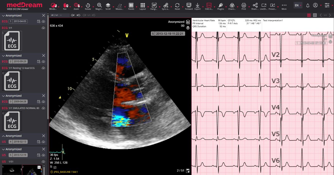


The MedDream DICOM Viewer provides not only standard image manipulation tools, but also a way to read, manipulate and interpret electrocardiography (ECG) DICOM data.
DICOM ECG Viewer’s manipulation tools are all presented in an innovative zoom model, which allows zooming in/out, measure and quantify the ECG data:

Measurement (mV, s). Area calculation indicating beats per minute, time, millivolt (mV, s, bpm);

QT points (RR, QT, QTc). QT interval - the RR interval is calculated as well as QT and the QTc (based on Bazett’s formula);

HR. Measure heart rate (HR) and compare its interval variance over the ECG;

QRS axis. Measure the QRS electrical heart axis;

Studies comparison. Compare of two or more ECGs by normalizing and then overlaying them on one another.
DICOM ECG Viewer’s supported ECG devices:
Other parties’ brand names, with ownerships belonging to Mortara Instrument, custo med GmbH, SCHILLER AG, GE Healthcare, are not authorized by, sponsored by or associated with the MedDream trademark owner.
MedDream DICOM ECG Viewer can be to measure the volume of a 2D image by using Simpson’s approximation rule; the 2D area spun around a selected axis to form a 3D shape and the volume is measured. This technique allows volume measurements of a heart in a 2D computed radiography image.
The MedDream DICOM Viewer measurements specifically important for Cardiology are:

Volume. Volume measurement using circles created from a set number of points.

Cardiac volume. New method of measuring the Cardiac Volume.

CTR. Measure the cardiothoracic ratio (CTR) to estimate a heart size.
MedDream DICOM Viewer for Ultrasound supports standard image manipulation tools, a way to read, manipulate and interpret ultrasound (US) DICOM data including specific features for ultrasound data like the Velocity Time Integral (VTI) that can quantify the trace of the Doppler flow profile. MedDream DICOM Viewer features specifically important for Ultrasound are:

VTI (Velocity Time Integral). Used to measure the distance from which the blood was ejected over a date interval of time.

Velocity. One-click measurement that automatically displays the velocity value.

Up to 16 US studies may be opened at once.


This page covers Cardiology & Ultrasound features of MedDream DICOM Viewer. For other medical imaging fields, visit the main MedDream DICOM Viewer page or view the Specification file for full functionality.
What is the MedDream DICOM ECG Viewer?
+MedDream DICOM ECG Viewer is a specialized module within the MedDream platform designed specifically to read, manipulate, measure, and interpret electrocardiography (ECG) data in standard DICOM format directly via a web browser. MedDream’s web DICOM Viewer provides standard cardiology image manipulation tools and a way to read, manipulate and interpret electrocardiography data:
Does the viewer support cardiology and ECG files?
+Yes, MedDream features dedicated cardiology tools. It supports multi-frame cine-loop playback for ultrasound and offers a specialized ECG module for viewing, measuring (mV, s, bpm), and interpreting electrocardiography data.
What specific ECG measurements are supported?
+The viewer features dedicated measurement tools including Area calculation (mV, s, bpm), Heart Rate (HR) tracking with interval variance, QRS electrical heart axis measurement, and detailed QT point analysis (RR, QT, and QTc based on Bazett’s formula).
Which ECG device manufacturers does MedDream support?
+MedDream supports 12-lead DICOM ECG files (Little Endian) generated by major devices including Mortara, Custo med, Schiller, GE, and any other devices capable of producing standardized DICOM ECG data via a gateway.
Does the viewer support Cardiology Ultrasound (US) studies?
+Yes, the MedDream viewer natively supports Cardiology Ultrasound and Intravascular Ultrasound (IVUS) studies. It features multi-frame cine-loop playback essential for echocardiography review. MedDream’s web DICOM viewer supports ultrasound (US) and intravascular ultrasound (IVUS) studies with VTI measurement and multi-frame cine-loop playback:
Can MedDream measure Cardiac Volume?
+Yes, the viewer includes an advanced tool to measure the volume of a 2D image using Simpson’s approximation rule. This allows cardiologists to spin a 2D area around a selected axis to calculate the volume of the heart from a 2D computed radiography image.
Is there a tool for Doppler flow profiles?
+Yes, MedDream supports the measurement of the Velocity Time Integral (VTI) on ultrasound studies. This specialized tool is used to quantify the trace of the Doppler flow profile.
Are standard image manipulation tools available for cardiology?
+Yes, standard tools such as zoom, pan, windowing, and precise scaling are fully available and presented in an innovative zoom model optimized for visualizing high-frequency ECG waveforms.
Does it require local installation to view ECGs?
+No, the ECG module is part of the HTML5 zero-footprint MedDream viewer. Cardiologists can access and measure ECGs securely from any desktop or mobile device with a modern web browser.
Can MedDream integrate with Cardiology Information Systems?
+Yes, through its open URL, Communication API, and Viewport API, MedDream can be seamlessly embedded into third-party CVIS, HIS, or EHR platforms to display ECGs seamlessly within the patient’s record.
Does the web DICOM viewer support ECG and Ultrasound measurements?
+MedDream supports specific measurements for ECG and Ultrasound. For Ultrasound, it supports Velocity Time Integral (VTI) measurements to quantify Doppler flow profiles and multi-frame cine-loop playback. For ECG data, it features an innovative zoom model that calculates Area (mV, s, bpm), QT/QTc points, Heart Rate (HR), and QRS electrical heart axis.
Is the ECG viewer module certified?
+Yes, the cardiology and ECG viewing functionalities are covered under MedDream’s overarching FDA 510(k) clearance and CE Class IIb medical device certification for primary diagnostic use.

© 2026 MedDream UAB. All rights reserved.


 Software vendors
 Hardware vendors
 System Integrators
 AI & CAD vendors
 Education & Research
 AWS HealthImaging
 Azure Health Data Services
 Google Cloud Healthcare API
 Radiology
 Cardiology & Ultrasound
 Ophthalmology
 Orthopedics
 Dentistry
 Surgery
 Mammography
 Pathology
 Veterinary


